ICCS Portland Plenary session III: International Focus. Chair: Mariela Monreal.
The theme for the afternoon plenary session on Monday (October 17th 2011) was “international focus”. The session was chaired by Dr. Mariela Monreal (citometria@fundaleu.org.ar) of FUNDALEU (Foundation to Fight Leukemia), Buenos Aires, Argentina. Four international speakers presented their work related to the use of flow cytometry in diverse areas of hematological and lymphoid abnormalities.
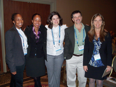
International Focus Plenary Session presenters (from left to right) Ms Songee Branch, Dr. Kim Quimby, Dr. Elaine Costa, Dr. Jorge Rossi, and Dr. Mariela Monreal (session chair).
The first speaker was Elaine Costa, MD (elainesc@centroin.com.br) who is a pediatrician at the Pediatrics Institute IPPMG, Federal University of Rio de Janeiro (UFRJ), Rio de Janeiro, Brazil. She presented the “B-cell maturation patterns in normal and regenerating bone marrow using the BCP-ALL Euroflow panel”. She started her presentation by outlining the classical scheme of B-cell differentiation and emphasized the importance of the normal pattern of antigen expression in detection of minimal residual disease in precursor B-cell neoplasms. Next she described the composition of the standardized EuroFlow four tube, eight color antibody panel used for B-cell precursor acute lymphoblastic leukemia diagnosis, the BCP-ALL panel, tested in 148 leukemic samples. (Details of this and other EuroFlow panels are at http://www.euroflow.org/imagenes/news/euroflow_handout_on_antibody_panels.pdf). She emphasized that the traditional visualization of flow cytometry data by pairwise combinations of antibodies creates an unacceptably large number of scattergrams and a comprehensive analysis of the precise antigen expression pattern may not occur in this traditional approach. The solution to this difficulty is the use of principal component analysis (PCA) of a unified data file with a potentially infinite number of dimensions (Cytometry A. 2008;73:834-46). Dr. Costa has previously used this approach to subclassify mature B-cell neoplasms (Leukemia. 2010;24:1927-33). In this approach, each event from the first tube is “marked” using three or more anchoring parameters (forward scatter, side scatter and CD19). In the subsequent tubes, a “nearest neighbor” is assigned for each event in the first tube based on the closeness of values of the anchoring parameters in the subsequent tubes. The antigen expression values for these assigned events are then added to the directly measured antigen expression values for the anchoring event in the first tube. This is accomplished by using INFINICYT software (Cytognos SL, Salamanca, Spain). This merged “list mode” data file contains the antigen expression value (directly measured or assigned) for every antigens tested in the panel for every event in the first tube. Afterward, Principle Component Analysis is applied to data from a group of 10 or so patients with the same condition and graphically visualized through the automated population separator (APS) view of the INFINICYT software. A clearly identifiable pattern is seen for different B-cell neoplasms using this approach in almost every case. Then Dr. Costa presented data from her recent studies in which she has applied the same approach to 33 bone marrows from patients treated for B-ALL. She successfully identified minimal residual disease and differentiated it from normal regenerating marrow elements in these samples by the automated approach. This study illustrated the potential for automation if a standardized approach is used for initial staining as advocated by EuroFlow. The downside of the approach is the rather large number of antibodies needed for the analysis. Traditional flow cytometry users may also be unsettled by the fact that highly derivative data are being used to make potentially critical patient management decisions. More experience with these automated approached may relieve these apprehensions if the findings are robustly repeated.
The second presentation was given by Kim Quimby, MBBS (kim.quimby@cavehill.uwi.edu) who is a young physician from the Chronic Disease Research Centre, Tropical Medicine Research Institute in Bridgetown, Barbados and is currently pursuing a PhD in immunology at the University of West Indies Tropical Medicine Research Institute. She spoke about the phenotypic commitment of monocytes towards a protective hemoglobin scavenging phenotype (CD14posCD163highHLA-DRlow) under hemolytic conditions. Intravascular hemolysis may cause tissue injury directly or via a systemic inflammatory response. Under physiological conditions, free hemoglobin released by hemolysis is bound by haptoglobin and the complex is internalized via the hemoglobin scavenger receptor CD163 on monocytes. This facilitates the catabolism of hemoglobin via heme-oxygenase-1. Recently, a novel subset of CD68pos/ CD163high/ HLA-DRlow macrophages with high expression of heme oxygenase-1 was recognized in hemorrhagic areas of atherosclerotic plaques. Red blood cells in patients undergoing cardiopulmonary bypass undergo lysis to variable degree during the procedure. Ms Quimby hypothesized that the hemolysis would lead to a compensatory evolution of a CD163high/ HLA-DRlow phenotype in circulating CD14pos monocytes after such surgery. Using three color flow cytometry, she examined the phenotype of circulating monocytes in 12 patients before and after surgery requiring bypass. In additions, the concentration of haptoglobin – hemoglobin complexes in the serum was measured. A striking phenotypic switch from the preoperative phenotype of CD163low/ HLA-DRhigh monocytes occurred to CD163high/ HLA-DRlow phenotype 24 hours after the surgery. At the same time the intracellular concentration of heme oxygenase-1 also went up significantly. These phenotypic changes were preceded by peak Hemoglobin-Haptoglobin levels observed at 2 hours after surgery, which suggests a plausible causal link between the hemolytic trigger for the phenotypic changes. After describing this previously published work (Cytometry Part B (Clinical Cytometry). 2010; 78B:357–360) Dr. Quimby described newer results from sickle cell patients (Br J Haematol. 2011;153 (supplement 1):40). Sickle cell disease is a significant public health issue in Barbados and it is of interest to know if the hemolysis associated with sickle cell disease also produces the protective phenotype in monocytes. To their surprise they found that the circulating monocytes in sickle cell patients have lower expression of CD163 than controls. This was explained in part by their observation that serum haptoglobin levels were low in these patients suggesting chronic hemolysis may have depleted haptoglobin. The lack of stimulatory signals by the hemoglobin-haptoglobin complex may underlie the low CD163 expression in these patients.
After the coffee break, Dr. Jorge Rossi (jrossi@garrahan.gov.ar) from Hospital Nacional de Pediatria, Argentina, gave the third presentation on characterization of pediatric acute leukemias of unusual immunophenotypes. Garrahan Hospital has 510 beds and receives over 370 new children with malignant diseases every year as it is a referral institution in the region. He has analyzed the immunophenotypes of over 1500 cases seen over about 17 years. From this pool three groups of unusual immunophenotype were identified: (a) 3 cases of plasmacytoid dendritic cell leukemia, (b) 9 cases which showed complete lineage switch after treatment, and (c) 64 cases with ambiguous lineage (considering EGIL scoring system). The 3 cases of plasmacytoid dendritic cell leukemia were reported earlier (Leuk Lymphoma. 2006; 47:715-25) and were from a total 1363 new patients with acute leukemia diagnosis. The leukemic blasts had a lymphoblastic morphology (i.e. FAB L2). They lacked the most specific markers for myeloid, T or B lineage and CD34. The cells expressed CD4, CD56, HLA-DR, BDCA-2 (CD303) and BDCA-4 (CD304). Skin involvement was not seen in any patient, unlike adult patients with this entity, and all showed good response to acute lymphoblastic leukemia (ALL) protocols, achieving complete remission even when one of the patients relapsed and received an allogeneic transplant. The 9 cases of lineage conversion included 7 ALL cases converting to AML and 2 converting in the opposite direction. All except one patient were infants, the sole exception being an adolescent. The switch occurred as early as 8 days after initiating chemotherapy to a few months. Importantly, cytogenetic and/ or molecular studies in these patients confirmed that the leukemic clone was the same as before but the antigen expression pattern had completely changed. Seven of the 9 cases had cytogenetic abnormalities involving the MLL gene. Dr. Rossi discussed the modern concept of plasticity in maturation pathways and the potential for cross over and reversal of certain maturation sequences. Lastly he discussed the revised definition of leukemias of ambiguous lineage in the WHO (2008) classification. By applying these stricter criteria for lineage assignment, the originally identified number of 64 apparent ambiguous lineage acute leukemia cases shrunk to 35 cases (2.54% of total). These included several cases of B/myeloid and T/myeloid biphenotypic and one case of tri-phenotypic leukemia. Bilineal leukemia was occasionally seen and undifferentiated leukemias were also rare. Interestingly several rare combinations were noted, such as megakaryoblastic/ T-ALL which showed cytogenetic abnormality of 14q32 and responded to AML chemotherapy. MLL abnormalities and chromosome 7 abnormalities were frequently observed in these patients. This group of patients presented poorer response to treatment and outcome than that of classical ALL and AML cases.
The last speaker in this session, Mrs. Songee Branch-Beckles (Clinical Care & Infrastructure Management Preceptee) is the Clinical Information Specialist for the Ladymeade Reference Unit, the national HIV/AIDS treatment center in Barbados. In this capacity, Mrs Branch-Beckles is responsible for the day to day running of the laboratory and ensuring the quality of lab services rendered to approximately 1400 patients with HIV annually. She has completed studies in Medical Laboratory Technology at Barbados Community College and post graduate studies at the University of the West Indies, Cave Hill campus and Michigan State University, respectively. She is currently enrolled as a PhD student at the University of West Indies. She presented her work of standardizing a two color protocol using pan-leuko-gate (PLG protocol) for CD4 count instead of the more prevalent but expensive four color protocol. She prefaced her talk with the dire statistics about HIV in the Caribbean nations, which have the world’s second highest HIV infection rate. In Barbados CD4 counting was sporadically available since the 1990s but escalating costs had deprived most needy patients of this basic test required for proper disease management. The national government of Barbados established a comprehensive care center for HIV patients called the Ladymeade Reference Unit earlier in this decade which has transformed HIV care in the country. Since cost containment is critical in order to achieve universal care for HIV patients in a resource limited setting, the Ladymeade Unit has tested a two color flow cytometry protocol using only CD45-FITC and CD4-PE antibodies as published previously (Cytometry B Clin Cytom. 2008;74 Suppl 1:S40-51). They directly compared the results of the two color protocol from 153 samples with the results of the more widely accepted 4 color protocol to validate this simpler and economical protocol. They found the correlation with the standard 4 color protocol, both for CD4 percentages and absolute counts to be very good, with very little bias throughout the range of values. Importantly, the cost of the test was reduced from $26 to $8 per sample. This has reduced the yearly expense for testing from $119,000 to $52,000, thus freeing up $67,000 for treatment of the 1500+ patients cared for by the Unit.
This session emphasized the scope of international work in clinical flow cytometry, from basic testing appropriate for the circumstances to the most advanced algorithmic and highly sophisticated analysis using a large panel of antibodies on multicolor platforms. The enthusiasm and sense of purpose was palpable in each of the presenters. Their presentations enriched the experience of the US audience as well.

Anand Lagoo, MD, PhD
Duke University, Durham, NC