Ask an Expert
Back to Categories
How are basophils identified by flow cytometry?
Basophils are the least common of the normal circulating leukocytes, usually representing less than 1% on a peripheral blood differential count. Basophils are part of the innate immune system and are thought to participate in defense against parasites. They also play a role in allergic inflammatory reactions. Basophils can be increased in some myeloproliferative disorders, such as chronic myelogenous leukemia.
Basophils have distinct immunophenotypic features by flow cytometry. They are in the dim CD45/low side scatter area or "blast" gate (figure 1). There are different cell types in the "blast" gate: progenitors, myeloid blasts, degranulated myeloid cells, hematogones, plasmacytoid dendritic cells (pDCs), and basophils. It is important to recognize these cells especially in doing minimal residual disease assays for leukemia so an abnormal blast population is not confused for other normal cell types.

Figure 1: "Blast" gate (red) in the CD45 vs. SSC (linear) and CD45 vs. SSC (log) dot plots
Both basophils and pDCs express bright CD123 and this marker can be used to differentiate them from other cells. However, basophils can be separated from pDCs by the absence of HLA-DR expression (Figure 2).
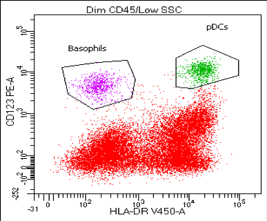
Figure 2: Basophils (purple) vs. pDCs (green) in the "blast" gate (dim CD45/low side scatter)
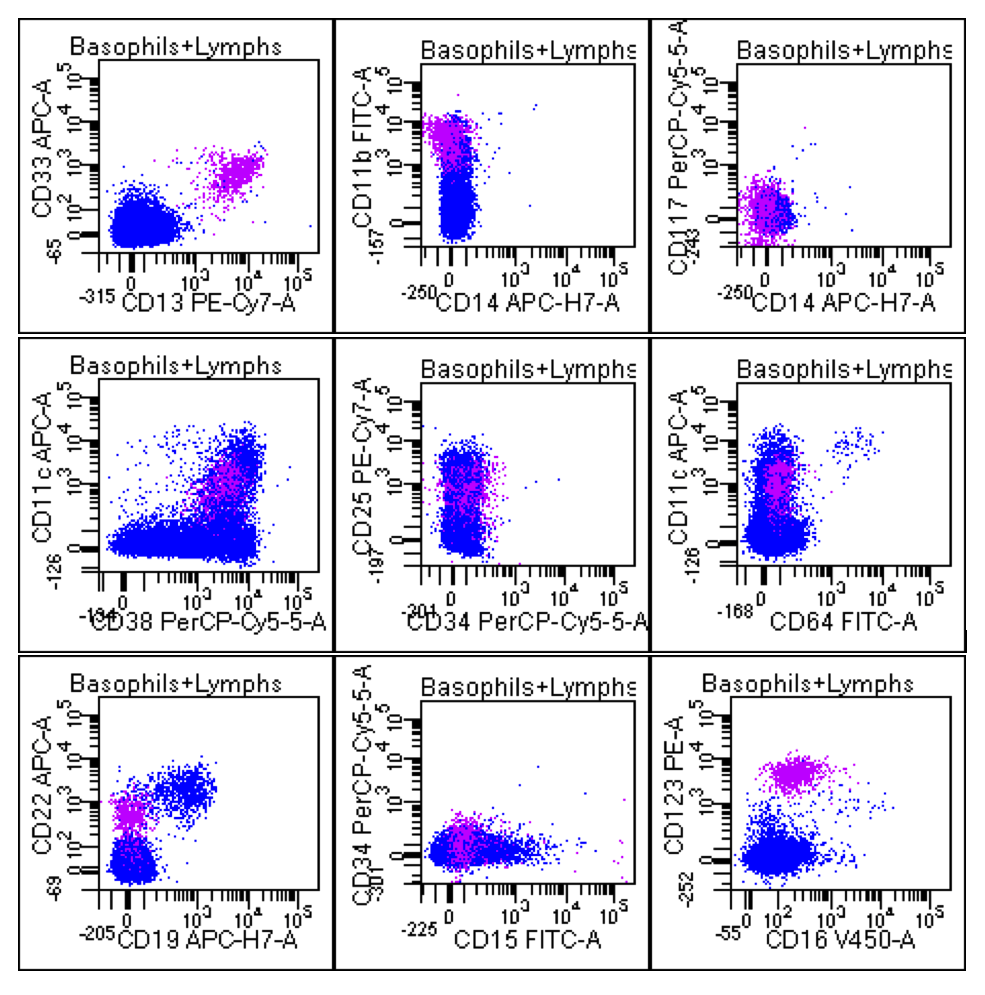
In addition, basophils express CD11b, dim CD11c, CD13, dim CD22, dim CD25, CD33, and bright CD38. They lack the expression of CD14, CD15, CD16, CD19, CD34, CD64, and CD117 . The dot plots below show the basophil population in purple in comparison with the lymphocyte population in blue.
References:
Han X, Jorgensen JL, Brahmandam A, et al. (2008) Immunophenotypic study of basophils by multiparameter flow cytometry. Arch Pathol Lab Med. 132:813-819.
Author: Kim Le