What is the sequential gating strategy to select singlets, remove debris?
The standard initial sequential gating strategy in clinical flow cytometry involves: 1) time gating, 2) singlet gating (doublet exclusion), 3) viability gating (fs/ss and/or viability dye), and 4) CD45/SS gating.
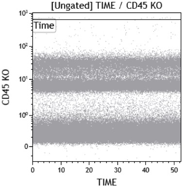
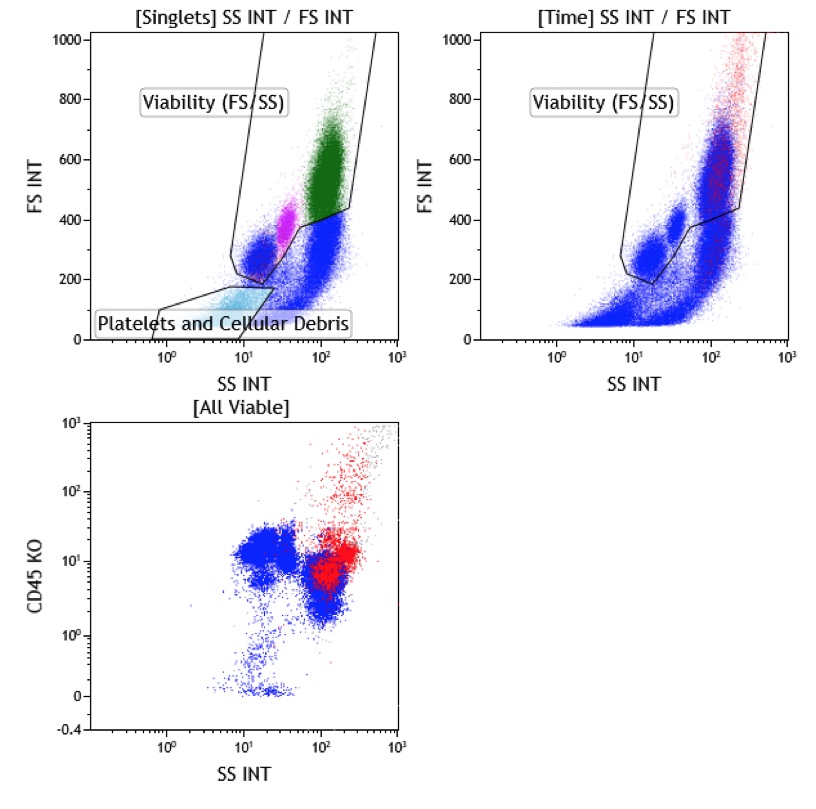
1. Time gating is used to exclude disruption of the flow of events due to clots, clogs and bubbles (i.e. fluidics issues) which can give rise to spurious events. In the second plot, notice the drop off of CD45 intermediate events near the end of the collection which could lead to spurious undercounts or overcounts of differing populations. Left shows an example of uninterrupted collection. Right shows an example of air aspiration at the end of collection.
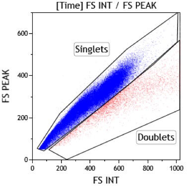
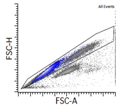
2. Singlet gating (or doublet exclusion) uses forward light scatter characteristics to remove events that are likely doublets (two cells interrogated simultaneously). Exclusion of these events is necessary as they may introduce false cells populations. Note however some real singlet events do fall in the doublet region, such as proliferative and/or hyperdiploid cells (e.g. neoplastic plasma cells).
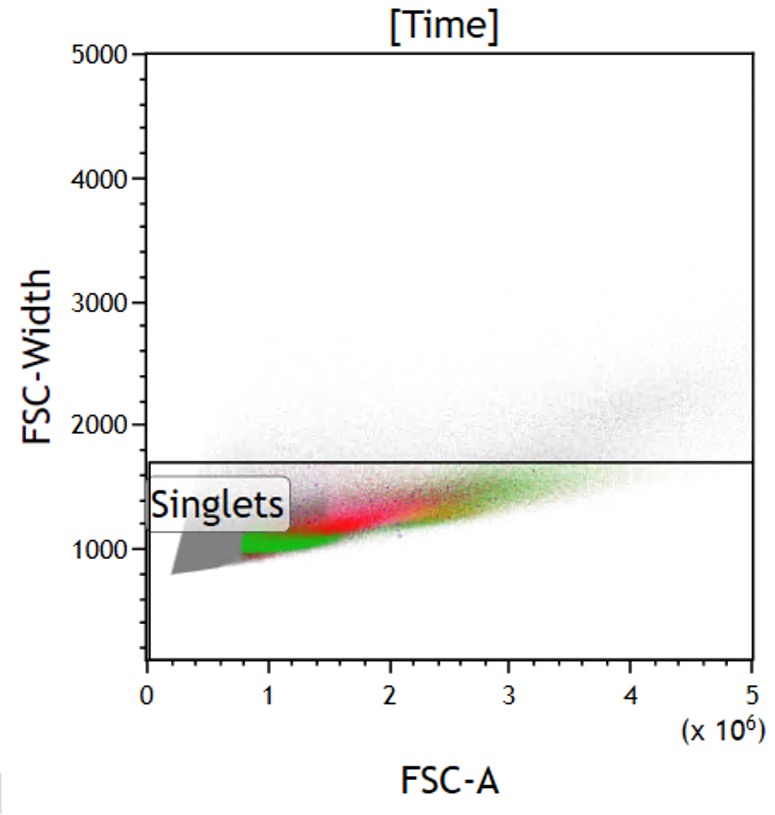
An alternative method for singlet gating uses the increased width of the forward scatter signal among double events and shows similar results.
3. After doublet exclusion, a viability gated based on FS/SS is used. Typically, the smallest cells of interests are lymphocytes (or erythroid precursors depending on the quality and type of lysis used) and could be the smallest cell type by FS included in the FS/SS viability gate. Of note, events with low side scatter and forward scatter (colored light blue) are typically platelets and cellular debris which often bind antibodies nonspecifically. Also note if the doublets (colored red) were not removed as in the previous step (colored red), they will be included with high FS/SS events which may also bind nonspecifically or contain disparate cellular phenotypes on other plots (CD45/SS seen here).
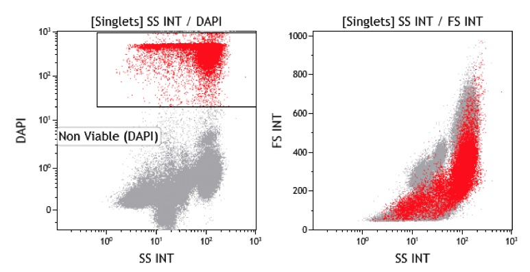
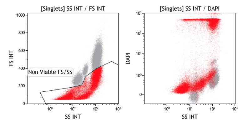
In addition to FS/SS based viability gating, several viability dyes are commercially available which either differentially bind dead/permeabilized or live/metabolically active cells. One such dye displayed below, 4?,6-diamidino-2-phenylindole (DAPI) will bind intracellular DNA on dead/permeablized cells. These dead cells (gated in red) may or may not bind nonspecifically and so are excluded in analysis. Ideally, viable cells should not show any signal, however, it is important to know cellular debris can be also negative (perhaps due to lack or minimal amount of nuclei acids) limiting the specificity of this strategy. Another consideration with the viability dye is the spectral interference with other fluorochromes due to their typically wide emission spectra although the effect is somewhat lessened since we aim exclude the offending positive events. Finally, the addition of a viability dye decreases the number of markers that can be tested in a panel. Some labs use a separate tube with a viability dye in addition to FS/SS to help delineate viable cells (i.e. backgating) based on FS/SS while other labs will include a viability dye in some of their screening panels.
Note that while FS/SS and DAPI will exclude similar populations (i.e. overlap), there are also populations selected by one methods that are not included by the other and therefore a combined approach using BOTH FS/SS viability and a viability marker should be considered to minimize non-viable events in a specimens.
Common commercially available viability dyes in flow cytometry include DAPI, PI, 7-AAD, Syto16, etc. DAPI, PI and 7-AAD are membrane impermeable dyes binding to nucleic acids and therefore cannot be used to exclude non-viable events in the setting of membrane permiablitization (e.g. for cytoplasmic and nuclear staining). Indeed, with the use of permeablization, these dyes are often used for detecting DNA content for cell cycle or ploidy analysis.
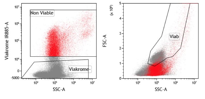
Alternative viability dyes are also available (often times called fixable dyes) which bind strongly to proteins in dead cells based on loss of membrane integrity and can be used in the setting of membrane permeablization. The example below shows one such dye (viakrome 808 gated below in red) being excluded by live cells, as seen in this markedly degenerated specimen. However note that there are a plethora of commercially available dyes for almost any conceivable laser/filter combination.
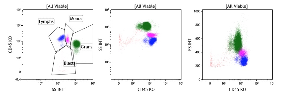
4. Finally, leukocyte separation based on the classic CD45/SS gating can be performed.
Author:
David Ng, MD
University of Utah
Reviewed by:
Jolene Cardinali Huifei Liu Bakul Dalal