Ask an Expert
Back to Categories
What Are CD11b And CD11c and What Are Their Utility in The Clinical Flow Cytometry Assessment For Hematologic Neoplasms?
CD11b, also known as integrin alpha M (ITGAM), is a glycoprotein that forms a heterodimer with CD18, to form Mac-1 (macrophage-1 antigen)1. CD11b is expressed in monocytes, macrophages, granulocytes (such as neutrophils) and most NK cells. The intensity of expression of CD11b varies at different stages of maturation of the neutrophils and monocytes. CD11b is negative on the early myeloid precursors. Its expression begins at the myelocyte stage, and the intensity of expression increases with mature neutrophils showing the highest intensity of CD11b. Eosinophils and basophils express CD11b2.

Figure 1: A) CD11b expression on the granulocytes in different stages of maturation with the brightest expression on neutrophils. B) CD11b positive eosinophils C&D) Basophils showing CD11b expression.
In the monocytic lineage, CD11b is absent on the monoblasts. Promonocytes begin to express CD11b and is brighter on mature monocytes. Mature B and T-lymphocytes are generally negative for CD11b. A subset of reactive T-cells and T-LGL leukemia are shown to express CD11b2.
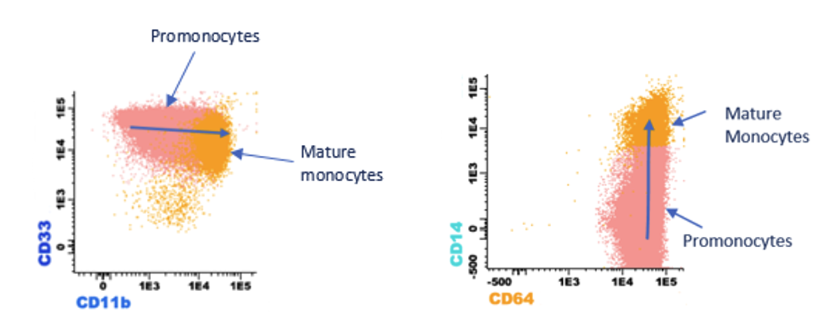
Figure 2: CD11b expression in the monocytic lineage in different stages of maturation with the brightest expression on mature monocytes
CD11c, also known as integrin alpha X (ITGAX), is a glycoprotein that forms a heterodimer with CD18 to form complement receptor 4 (CR4)1. CD11c is positive on monocytes, macrophages, granulocytes and the subset of lymphocytes such as activated T-cells and NK cells2. However, the intensity of expression varies among different cell types, with monocytes showing the strongest CD11c expression and neutrophils and lymphocytes showing lower levels of CD11c expression. In the granulocytes, CD11c expression is seen first at the myelocyte stage and is usually dim. CD11c is brighter on mature neutrophils2. Eosinophils are also positive for CD11c. In the monocytic lineage, CD11c is positive on the monoblasts, with the intensity increasing as the cells mature. The strongest CD11c expression is seen on mature monocytes.

Figure 3: CD11c expression in granulocytes, monocytes and NK cells
Utility of CD11b and CD11c in hematologic neoplasms
CD11b
Acute myeloid leukemia (AML) with monocytic differentiation is usually positive for CD11b3. Blasts of untreated acute promyelocytic leukemia (APL) are often negative for CD11b and CD11c although can acquire CD11b and CD11c following treatment with arsenic and ATRA due to differentiation4,5,6. Myeloid blasts of AML occasionally show aberrant expression of CD11b and is often associated with a monosomal karyotype and poor prognosis7. Absence of CD11b and CD13 helps differentiate transient abnormal myelopoiesis (TAM) from AML in Down’s syndrome, which is usually CD11b and CD13 positive8. Expression of CD11b on granulocytes is decreased in myelodysplastic syndrome resulting in the loss of the convex pattern of granulocyte maturation pattern in CD11b Vs CD169.
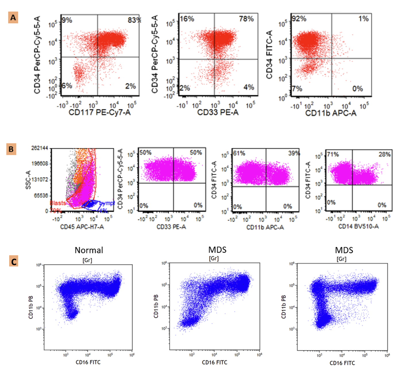
Figure 4: A) CD11b negative myeloid blasts (positive for CD34, CD117 and CD33). B) Monoblasts showing heterogenous expression of CD11b. C) Abnormal maturation pattern of dysplastic granulocytes in CD11b Vs CD16 in MDS patients (Image credit: Cara Shirai, Washington University in St. Louis).
In B-lymphoblastic leukemia/lymphoma (B-ALL/LBL), CD11b expression can help identify measurable residual disease (MRD). The blasts may show progressively increased intensity of CD11b expression and therefore help distinguish neoplastic blasts from hematogones, which are dim to negative10. CD11b expression is also shown to predict therapy resistance in B-ALL10.
CD11c
CD11c is one of the monocytic differentiation markers. Blasts of AML are variably positive for CD11c2,3. Further, within lymphoid neoplasms, CD11c is positive on hairy cell leukemia, splenic B-cell leukemia/lymphoma with prominent nucleoli (previously hairy cell leukemia variant) and a subset of splenic marginal zone lymphoma3. CD11c is shown to have some utility in differentiating chronic lymphocytic leukemia (CLL) from mantle cell lymphoma (MCL). CLL may show expression of CD11c while MCL is almost always negative for CD11c11.

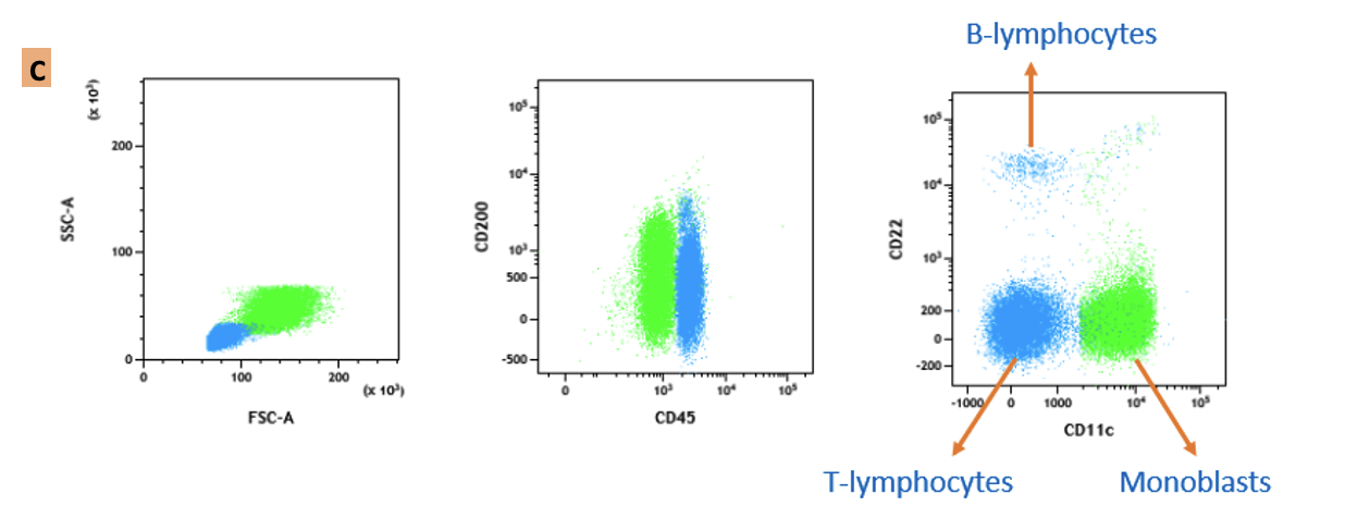
Figure 5: CD11c positive neoplastic B-cells in A) hairy cell leukemia and B) splenic B-cell leukemia/lymphoma with prominent nucleoli hairy cell leukemia variant); C) CD11c expression on monoblasts (Image credit: George Deeb, Emory University school of Medicine.
References
1. Mazzone A, Ricevuti G. Leukocyte CD11/CD18 integrins: Biological and clinical relevance. Hematologica 1995; 80:161-175.
2. Gorczyca W. Flow cytometry in neoplastic hematopathology – morphologic-immunophenotypic correlation. 3rd ed. CRC press; 2017.
3. Swerdlow SH, Campo E, Harris NL, Jaffe ES, Pileri SA, Stein H, Thiele J (Eds.): World Health Organization classification of Tumours of Haematopoietic and Lymphoid Tissues. Revised 4th ed. Lyon: IARC; 2017.
4. Zhang T, Westervelt P, Hess JL. Pathologic, cytogenetic and molecular assessment of acute promyelocytic leukemia patients treated with arsenic trioxide (As2O3). Mod Pathol. 2000 Sep;13(9):954-61. doi: 10.1038/modpathol.3880174. PMID: 11007035.
5. Tohyama K, Shiga S, Fujimoto H, Hamaguchi Y, Ichiyama S. Automated analysis of differentiation-induced leukemic cells during all-trans retinoic Acid therapy of acute promyelocytic leukemia. Arch Pathol Lab Med. 2003 Jan;127(1):e4-10. doi: 10.5858/2003-127-e4-AAODIL. PMID: 12562284.
6. Horna P, Zhang L, Sotomayor EM, Lancet JE, Moscinski LC. Diagnostic immunophenotype of acute promyelocytic leukemia before and early during therapy with all-trans retinoic acid. Am J Clin Pathol. 2014 Oct;142(4):546-52. doi: 10.1309/AJCPPOKEHBP53ZHV. PMID: 25239423; PMCID: PMC5744863.
7. Chen MH, Atenafu E, Craddock KJ, Brandwein J, Chang H. CD11b expression correlates with monosomal karyotype and predicts an extremely poor prognosis in cytogenetically unfavorable acute myeloid leukemia. Leuk Res. 2013 Feb;37(2):122-8. doi: 10.1016/j.leukres.2012.09.019. Epub 2012 Oct 23. PMID: 23092917.
8. Karandikar NJ, Aquino DB, McKenna RW, Kroft SH. Transient myeloproliferative disorder and acute myeloid leukemia in Down syndrome. An immunophenotypic analysis. Am J Clin Pathol. 2001 Aug;116(2):204-10. doi: 10.1309/XREF-C9T2-6U0A-4EDT. PMID: 11488066.
9. Chung JW, Park CJ, Cha CH, Cho YU, Jang S, Chi HS, Seo EJ, Lee JH, Lee JH, Lee KH, Im HJ, Seo JJ. A combination of CD15/CD10, CD64/CD33, CD16/CD13 or CD11b flow cytometric granulocyte panels is sensitive and specific for diagnosis of myelodysplastic syndrome. Ann Clin Lab Sci. 2012 Summer;42(3):271-80. PMID: 22964615.
10. Rhein P, Mitlohner R, Basso G, Gaipa G, Dworzak MN, Kirschner-Schwabe R, Hagemeier C, Stanulla M, Schrappe M, Ludwig WD, Karawajew L, Ratei R. CD11b is a therapy resistance- and minimal residual disease-specific marker in precursor B-cell acute lymphoblastic leukemia. Blood. 2010 May 6;115(18):3763-71. doi: 10.1182/blood-2009-10-247585. Epub 2010 Mar 12. PMID: 20228269.
11. Kraus TS, Sillings CN, Saxe DF, Li S, Jaye DL. The role of CD11c expression in the diagnosis of mantle cell lymphoma. Am J Clin Pathol. 2010 Aug;134(2):271-7. doi: 10.1309/AJCPOGCI3DAXVUMI. PMID: 20660331.
Author: Rajeswari Jayakumar, Cara Shirai and George Deeb