
Ask an Expert
Back to Categories
PNH Testing Review
UPDATES IN PNH TESTING
Flow Cytometric evaluation for Paroxysmal Nocturnal Hemoglobinuria (PNH) is currently considered the gold standard for the detection and quantification of PNH clones based on high-sensitivity WBC analysis and RBC analysis. Based on the ICCS/ESCCA PNH Guidelines the most tested WBC panels are FLAER/CD24/CD15/CD64/CD14/CD45 or FLAER/CD157/CD15/CD64/CD45 and the most tested RBC panel uses CD59 and CD235a with a recently recommended inclusion of CD71 (see examples below).
WHITE BLOOD CELL ASSAYS
Two GPI-linked structures need to be examined for each assessed WBC lineage. Combinations such as FLAER and CD24 (Figure 1), or FLAER and CD157 (Figure 2), are commonly used to detect GPI-deficient neutrophils. Similarly, FLAER paired with either CD14 (Figure 1) or CD157 (Figure 2) is the most validated combination for detecting GPI-deficient monocytes. Both assays below have worked well in laboratories performing PNH testing for the identification and quantification of both larger and minor PNH clones.
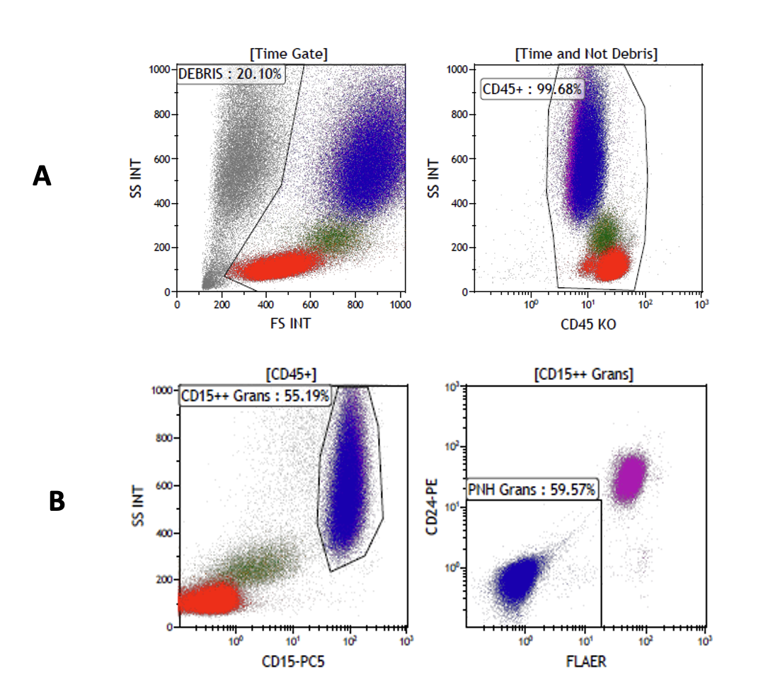
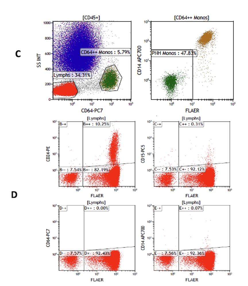
Figure 1: 6-color PNH WBC Panel FLAER/CD24/CD15/CD64/CD14/CD45
A. CD45+ cells are gated on “Time” with the exclusion of the Debris (“Time” and “Singlets” plots are not shown).
B. CD45+ cells are gated lineage-specifically on CD15++ neutrophils and show a FLAER/CD24 negative PNH clone within the neutrophil population.
C. CD45+ cells are gated lineage-specifically on CD64++ monocytes and show a FLAER/CD14 negative PNH clone within the monocyte population.
D. Lymphocytes are not suitable targets for PNH clone evaluation due to the long lifespan of memory cells but they do serve as excellent internal controls for instrument setup & compensation. All population should be visible on scale without under or over compensation and antibody performance can be verified (CD24+/FLAER+ B-cells, FLAER++/CD24-neg T-cells, FLAER int+/CD24 neg NK cells and FLAER-neg/CD24-neg PNH lymphocytes).
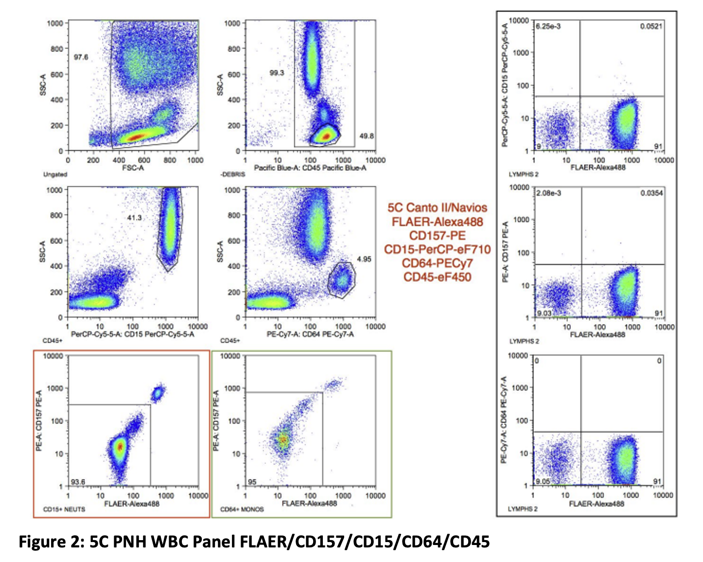
In this 5C panel (Figure 2), neutrophils are gated on CD45 and CD15 and monocytes are gated on CD45 and CD64. A PNH clone can be detected in both the neutrophils and monocytes based on the absence of the GPI markers FLAER and CD157. CD157 is highly expressed on both mature neutrophils and monocytes, allowing PNH WBC testing on cytometers with 5 or more PMTs. It is important to note that several CD157‐negative, non‐PNH cases have been observed in the authors’ laboratories. For these rare cases, the inclusion of the second GPI reagent (FLAER) as part of the built‐in robustness of the assay pre‐ vents the misinterpretation of the data as a PNH clone‐containing sample; the loss of two GPI‐linked structures is required to identify bona fide PNH phenotypes.
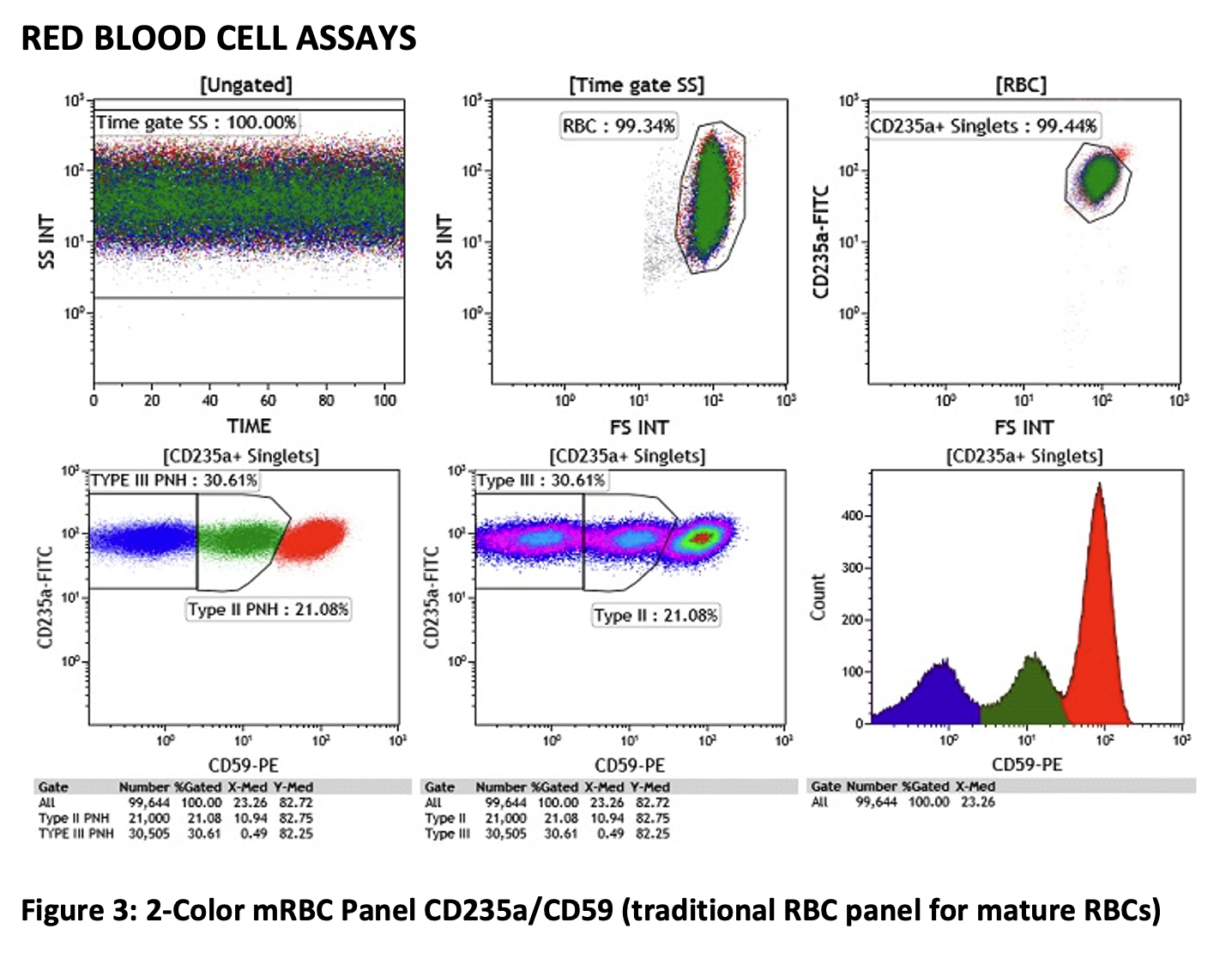
Figure 3 above shows the traditional 2C mature RBC (mRBC) assay. The top row shows the gating strategy with TIME and gating on singlets, while the bottom row shows a series of a dot plot, density plot as well as a single parameter CD59 histogram, which work well together for the purpose of finding the best region setting for separating PNH Type III cells (blue), PNH Type II cells (green) and normal RBCs (red).
Due to hemolysis of PNH RBCs and transfusions which "dilute" the PNH clone, the WBC assays are considered a more representative measure of “PNH clone size” than that derived from the RBC assay. However, the RBC assay is thought to be clinically useful for delineating Type III PNH phenotypes (total loss of CD59 expression) from Type II PNH phenotypes (partial loss of CD59 expression) and the Type II cells from Type I RBCs (normal CD59 expression). This information may be used to assess the effectiveness of complement blockade therapies.
Adding CD71: Interestingly, immature RBCs (iRBCs) are not hemolyzed by activated complement and are not significantly diluted by transfusions of mature RBCs. Therefore, some authors have proposed to assess immature RBC (nucleated RBCs and/or reticulocytes). More recently, antibodies targeting the transferrin receptor (CD71) have been utilized to detect immature RBCs (nucleated RBCs and reticulocytes).
- T• Old RBCs have been shown to express lower levels of CD59 than young RBCs and analysis of CD59 expression in mature RBCs in patients who have been receiving regular RBC transfusions will often show significant populations of apparent Type II phenotypes that are all derived from aging transfused normal Type I RBCs.
- • Also, looking at iRBCs, the CD71+ iRBC clone size is very close to clone sizes derived from neutrophils and/or monocytes.
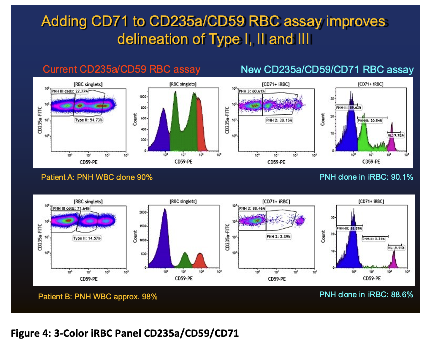
Figure 4 outlines the reasons for using the iRBC panel:
- The top row shows the RBC results for patient A, who has a WBC clone of 90%. The results of the mRBC assay (mature RBC assay shown in 2 plots on the left) shows a PNH type III clone (blue) and 2 populations (green) which appear to be PNH type II clones. Looking at the 2 plots to the right shows the iRBC assay which confirms the presence of 2 PNH type II populations. The total PNH clone in the iRBC is 90.1% which matches closely the PNH clone size in the WBC (90%).
- The bottom row shows the RBC results for patient B, who has a WBC clone of 98%. The results of the mRBC assay (mature RBC assay shown in 2 plots on the left) shows a PNH type III clone (blue) and a population (green) which appears to be a PNH type II clone. However, looking at the 2 plots to the right shows the iRBC assay which does not confirm the presence of a true PNH type II population. This is most likely due to transfused RBCs which often show decreased expression as they age. The PNH Type III clone in the iRBC is 88.6% which is slightly below PNH clone size in the WBC (98%).
SUMMARY
Most laboratories have experienced difficulties in the separation of the various PNH subsets from the normal RBCs. Part of this may be patient specific and the regions for the PNH type II, PNH Type III may have to be slightly adjusted to separate the PNH cells from the normal cells. However, it is often challenging or impossible to set the regions in order to accurately and reliably quantify the true PNH RBC clone size, especially for the PNH type cells. The addition of CD71 allows for confirmation of presence or absence of PNH Type II populations in the iRBCs in situations where misleading data are generated by monitoring only the mature RBC component. The extra cost of including CD71-APC to the regular two-color assay is minimal and given the higher quality of information that can be derived from the three-color version in PNH cases, we suggest that including CD71 would be a very useful assay modification.
REFERENCES:
- Sutherland DR, Richards SJ, Ortiz F, et al. CD71 improves delineation of PNH type III, PNH type II, and normal immature RBCS in patients with paroxysmal nocturnal hemoglobinuria. Cytometry. 2020;98:179–192. https://doi.org/10.1002/cyto.b.21853
- Sutherland, D. R., Illingworth, A., Marinov, I., Ortiz, F., Andreasen, J., Payne, D., … Keeney, M. (2018). ICCS/ESCCA consensus guidelines to detect GPI-deficient cells in paroxysmal nocturnal Hemoglobinuria (PNH) and related disorders: Part 2 - reagent selection and assay optimization for high sensitivity testing. Cytometry Part B, 94B, 23–48.
- Illingworth, A., Marinov, I., Sutherland, D. R., & Wagner Ballon, O. (2018). DelVecchio. ICCS/ESCCA consensus guidelines to detect GPI-deficient cells in paroxysmal nocturnal Hemoglobinuria (PNH) and related disorders: Part 3; data analysis, reporting and case studies. Cytometry Part B, 94B, 49–66.
- Oldaker, T., Whitby, L., Saber, M., Holden, J., Wallace, P. K., & Litwin, V. (2018). ICCS/ESCCA consensus guidelines to detect GPI-deficient cells in paroxysmal nocturnal Hemoglobinuria (PNH) and related disorders: Part 4 - assay validation and quality assurance. Cytometry Part B, 94B, 67–81.
- Borowitz, M. J., Craig, F., DiGiuseppe, J. A., Illingworth, A., Rosse, W., Sutherland, D. R., … Richards, S. J. (2010). On behalf of the clinical flow Cytometry society. Guidelines for the diagnosis and monitoring of paroxysmal nocturnal Hemoglobinuria and related disorders by flow Cytometry – A consensus guideline. Cytometry Clin Cytom, 72B, 211–230.
Author: Andrea Illingworth, MS, SCYM