
Ask an Expert
Back to Categories
When Should a Clonal B-Cell Population Be Classified as Monoclonal B-Cell Lymphocytosis or as Lymphoma? A Practical Flow-Based Approach
Monoclonal B-cell lymphocytosis (MBL) is defined as the presence of a clonal B-cell population in the peripheral blood at a count below the threshold for diagnosis of a lymphoproliferative disorder, in the absence of other features of a B-cell neoplasm. MBL is broadly classified into CLL-type MBL, which shares the immunophenotype of chronic lymphocytic leukemia, and non-CLL-type MBL, which does not. A further subgroup, atypical CLL-type MBL, has also been described, characterized by CD5 positivity but with bright CD20 expression, creating overlap with other CD5-positive B-cell neoplasms. Non-CLL-type MBL represents a clonal B-cell population that does not meet criteria for a defined lymphoma and does not demonstrate the classic CLL immunophenotype. Current NCCN guidelines emphasize that evaluation of small B-cell populations requires integration of flow cytometry with clinical findings, as flow cytometry alone is generally insufficient for definitive diagnosis [1,2].
Flow Cytometry Approach
Flow cytometry evaluation of peripheral blood typically focuses on CD5, CD10, CD19, CD20, CD23, CD11c, CD25, CD103, and kappa/lambda restriction, supplemented by CD200 when available. Some markers, such as CD5, CD10, CD20, and CD23, may be assessed by both flow cytometry and immunohistochemistry depending on specimen type and diagnostic context.
Interpretation Based on CD5 Status
Interpretation proceeds primarily based on CD5 expression, but the differential diagnosis is broader than a simple binary classification, particularly in CD5-positive disorders.
1. CD5-Positive Clonal B Cells
While the main differential diagnosis is between CLL-type processes and mantle cell lymphoma (MCL), atypical CLL-type MBL should also be considered in cases with CD5 positivity but non-classical CD20 intensity or borderline immunophenotypes.
|
Marker |
CLL-type (MBL/CLL) |
Atypical CLL-type MBL |
Mantle Cell Lymphoma (MCL) |
References |
|
CD5 |
Positive |
Positive |
Positive |
[1–2] |
|
CD20 |
Dim |
Bright |
Bright |
[1–2] |
|
CD23 |
Positive |
Variable/positive |
Negative/weak |
[1–2] |
|
Surface Ig |
Dim |
Bright |
Bright |
[1–2] |
|
CD200 |
Positive |
Positive or variable |
Negative/weak |
[3–6] |
|
FMC7 |
Negative |
Variable |
Positive |
[1–2] |
|
CD79b |
Negative/weak |
Variable |
Bright |
[1–2] |
|
Cyclin D1 |
Negative |
Negative |
Positive |
[1–2] |
Confirm MCL with:
• FISH for CCND1::IGH
• and/or Cyclin D1 immunohistochemistry
CD200 remains a useful discriminator, being strongly expressed in most CLL-type cases and typically absent or weak in MCL [3–6]. However, atypical CLL-type MBL may show intermediate CD20 and immunoglobulin intensity, which can partially overlap with other CD5-positive lymphoproliferative disorders and should not be interpreted in isolation.
FMC7 and CD79b remain useful adjunct markers in flow-based scoring systems such as the Matutes and modified Matutes scores, supporting classification of CLL-type immunophenotypes when interpreted in combination with CD23, CD5, and surface immunoglobulin intensity [1–2].
2. CD5-Negative Clonal B Cells
• CD10 positive → suggests follicular lymphoma [1]
• CD10 negative → broader differential including marginal zone lymphoma, lymphoplasmacytic lymphoma, and hairy cell leukemia
In CD5-negative small clonal populations, ancillary markers such as CD11c, CD25, CD103, and cytoplasmic immunoglobulin may help refine classification. However, these markers should be interpreted in context, as some entities (notably hairy cell leukemia) are rarely identified as incidental low-level peripheral blood clones, and their routine inclusion in all baseline screening panels may therefore be unnecessary outside specific clinical suspicion.
Clinical Criteria Essential for MBL Diagnosis (Per NCCN)
• Monoclonal B-cell count <5 × 10⁹/L
• No lymphadenopathy (nodes ≥1.5 cm)
• No splenomegaly
• No cytopenias attributable to marrow involvement
• No constitutional symptoms [2]
A complete blood count (CBC) with differential, including the absolute lymphocyte count (ALC), is essential and should be provided at the time of flow cytometric evaluation, as it is required for accurate classification of small B-cell clones and for application of diagnostic thresholds for MBL versus overt lymphoma.
When flow cytometry identifies a monoclonal B-cell population, the absolute monotypic B-cell count should be calculated using:
This value is critical for determining whether the clone meets criteria for CLL-type MBL (<5 × 10⁹/L in blood) or exceeds diagnostic thresholds for a lymphoproliferative disorder.
If systemic involvement is present, the peripheral blood findings should be interpreted as circulating lymphoma rather than MBL [2]. Thus, clinical evaluation, supported by CBC-derived absolute counts, remains the definitive discriminator between MBL and lymphoma.
Flow-Based Scoring Systems
The traditional Matutes score uses CD5, CD23, surface immunoglobulin, FMC7, and CD79b to distinguish CLL from other CD5-positive B-cell neoplasms [4,5]. However, modern studies demonstrate improved diagnostic accuracy when CD200 is incorporated into scoring algorithms [4–6]. Modified systems (e.g., CLL flow score) show increased specificity compared with the traditional Matutes score while maintaining high sensitivity [6].
• CD20 intensity
• Surface immunoglobulin intensity
• CD23 expression
• Cyclin D1 / t(11;14) testing when indicated
Case Example: Two MBL Populations in a Patient with CMML
Patient: 85-year-old male with chronic myelomonocytic leukemia under therapy
Specimen: Peripheral blood
CBC Findings
WBC: 3.16 K/µL
Hgb: 9.2 g/dL
Platelets: 17 K/µL
Differential
Neutrophils: 25.0%
Lymphocytes: 49.4%
Monocytes: 23.1%
Eosinophils: 0%
Basophils: 0.3%
IG: 2.2%
Morphology
Leukopenia with neutropenia, macrocytic anemia, and thrombocytopenia.
Gating Strategy:
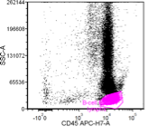
1. Lymphocytes are identified by bright CD45 expression with low side scatter.
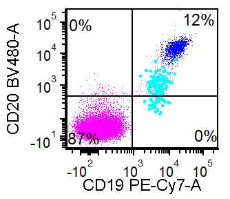
2. Gating on the lymphocytes, evaluation of CD19 expression, and in this case also CD20, enables identification of the B-cells. In this case 2 distinct B-cell populations are present based on level of CD20 expression and care colored as cobalt blue and teal.
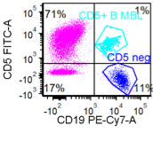
3. A separate tube of this sample was evaluated for T-cell markers in more detail, but evaluating for CD5 shows a discrete subset of B-cells with aberrant expression (turquoise), which corresponds to the subset showing dim to negative CD20.
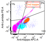
4. Coloring the populations allows their immunophenotypic features of each population to be evaluated including whether there is light chain restriction. Here the plot is gated on all lymphocytes to show their kappa and lambda light chain expression pattern. The CD5+ teal population is kappa-restricted, showing more dim light chain expression as compared to background lymphocytes. The cobalt population shows a cluster of B-cells with dim kappa expression, which is distinct from the abnormal CD5 population (turquose). Of note, there is also a polytypic B-cell population in the background with brighter light chain expression.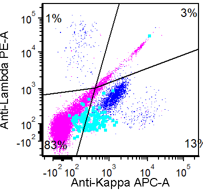
5. The subset of abnormal CD5-negative B-cells comprises the majority of B-cells in this CD5-negative, CD19+ gate, but the abnormal population can also be given a distinctive color (orange) to clarify their immunophenotypic pattern compared to background normal B-cells (remain cobalt).
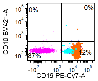
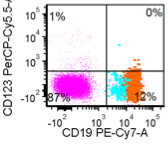
6. The immunophenotypes of the two abnormal B-cell populations can now be evaluated for the remaining markers tested. Both are negative for CD10 and CD123. The CD5+ subset (turquoise) shows slightly more dim expression of CD19 as compared to the CD5+ population (orange).
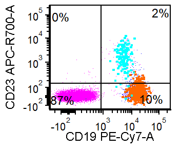
7. The CD5+ population (turquoise) expresses CD23 and slightly greater CD200 as compared to the kappa-restricted CD5-negative population.
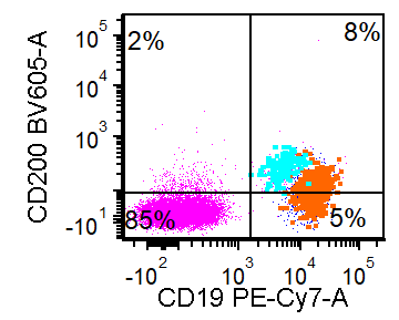
Summary of Flow Cytometry Findings
Two abnormal monoclonal B-cell populations identified:
1. Population 1 (turquoise): CD5+ population (~1% lymphocytes, 16 cells/µL): CD19+, CD20 dim to negative, CD5+, CD23+, CD200 dim, kappa restricted (very dim), negative for CD10 and CD123. → Consistent with CLL-type MBL
2. Population 2 (orange): CD5−/CD10− population (~12% lymphocytes, 190 cells/µL):
CD19+, CD20+, CD200 dim, kappa restricted
→ Non-specific phenotype, larger population
Background polytypic B-cells present. No abnormal T-cell population detected (not shown).
Conclusion
Flow cytometry is a critical component in the evaluation of mature B-cell lymphoproliferative disorders; however, it is not sufficient in isolation for definitive classification. Clinical staging remains essential in distinguishing monoclonal B-cell lymphocytosis (MBL) from overt lymphoma. In CD5-positive B-cell populations, mantle cell lymphoma (MCL) must always be excluded, using cyclin D1 immunohistochemistry and/or detection of t(11;14) by FISH. CD200 is a particularly valuable marker in differentiating chronic lymphocytic leukemia (CLL) from MCL. Overall, accurate diagnosis relies on an integrated approach that incorporates flow cytometry, ancillary studies, clinical context, and pattern recognition, which remains central in contemporary practice.
References
1. National Comprehensive Cancer Network. B-Cell Lymphomas. Updated 2026-03-12. https://www.nccn.org/professionals/physician_gls/pdf/b-cell.pdf
2. National Comprehensive Cancer Network. Chronic Lymphocytic Leukemia/Small Lymphocytic Lymphoma. Updated 2025-12-22. https://www.nccn.org/professionals/physician_gls/pdf/cll.pdf
3. Jain N, Wierda WG, O’Brien S. Chronic lymphocytic leukemia. Lancet. 2024;404:694–706.
4. Hoffmann J, Rother M, Kaiser U, et al. Determination of CD43 and CD200 improve B-cell lymphoma immunophenotyping. Cytometry B Clin Cytom. 2020;98:476–482.
5. Jalal SD. The contribution of CD200 to the diagnostic accuracy of Matutes score in the diagnosis of chronic lymphocytic leukemia in limited resources laboratories. PLoS One. 2021;16:e0247491.
Date published: September 21, 2026
Author: Salwa Khedr and Genevieve Crane