Ask an Expert
Back to Categories
How and in what situations should viability of specimens for flow cytometry be assessed?
The presence of non-viable cells in a specimen can be a serious issue in clinical flow cytometry. Non-viable cells can show non-specific binding of fluorescent antibodies or exhibit unusual autofluroescence that can result in the spurious identification of abnormal populations. In quantitative assays, such as those measuring T-cell subsets or CD34+ stem cells, the presence of non-viable cells can cause inaccuracy. Thus, the detection and exclusion of non-viable cells is an important part of flow cytometry analysis. However, the impact of viability varies across assays and specimens, and thus this analysis may be more important in some situations than others.
Viability is often specimen-dependent. Fresh blood specimens normally have excellent viability, but it decreases as the specimen ages. In fact, the CAP checklist for lymphocyte subset enumeration requires that labs demonstrate that their storage conditions preserve specimen integrity if stored longer than 24 hours. Assessment of viability is also required on stem cell products to ensure quantitative accuracy, but also because the viability may have an impact on engraftment.
For leukemia and lymphoma immunophenotyping, viability also often depends on the specimen. While it is generally high in fresh blood and bone marrow specimens, decreased cell survival is common in tissues and body fluids. This is particularly true in high-grade neoplasms with high turnover and specimens that have been exposed to significant processing or prolonged storage. For leukemia and lymphoma immunophenotyping, CAP regulations do not specify conditions that require viability testing. However, they do require that laboratories have a policy defining when viability assessment should occur.
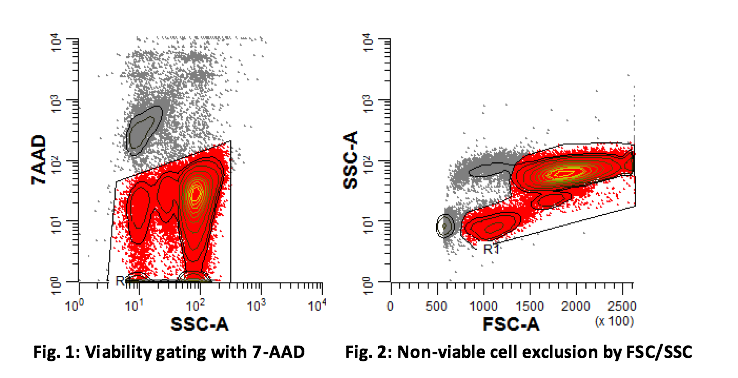
Assessment of viability is most accurately performed using a fluorescent viability dye. The most commonly used are propidium iodide (PI) and 7-aminoactinomycin D (7-AAD). Both of these dyes intercalate with DNA, so cellular staining requires a compromised membrane. Thus, only non-viable cells will be positive for these dyes. As a result, it is essential that staining occur prior to fixation, which causes membrane permeability. Both dyes are excited by a blue laser (488 nm) with emission maxima of 617 nm (PI) and 647 nm (7-AAD). Gating on viable events is performed by drawing a gate around the PI or 7-AAD-negative events on viability dye versus side scatter plot (see Fig. 1).
Because these dyes use up a slot in an antibody panel, it is not practical to utilize them in every panel. Instead, a rough viable cell gate can be drawn by excluding low-forward scatter (FSC) and high-side scatter (SSC) events (see Fig. 2). While this is not accurate for quantifying viability, it can be useful in excluding a large fraction of non-viable events to "clean up" immunophenotypic analysis.
Further Reading:
1. Clinical and Laboratory Standards Institute (CLSI). Clinical Applications of Flow Cytometry: Immunophenotyping of Leukemic Cells; Approved Guideline - Second Edition. CLSI Document H43-A2. 2007.
2. College of American Pathologists. Flow Cytometry Checklist. 2015.
3. Whitby A, Whitby L, Fletcher M, et al. (2012) ISHAGE protocol: are we doing it correctly? Cytometry B Clin Cytom. 82:9-17.
Author: Adam Seegmiller